What is a Foot Joint?
A solid understanding of anatomy is essential to effectively diagnose and treat patients with foot and ankle problems. Anatomy is a road map. Most structures in the foot are fairly superficial and can be easily palpated. Anatomical structures (tendons, bones, joints, etc) tend to hurt exactly where they are injured or inflamed. Therefore a basic understanding of surface anatomy allows the clinician to quickly establish the diagnosis or at least narrow the differential diagnosis. For those conditions that require a detailed understanding of anatomy is critical to ensure that the procedure is performed efficiently and without injuring any important structures. With a good grasp of foot anatomy it readily becomes apparent which surgical approaches can be used to access various areas of the foot and ankle.
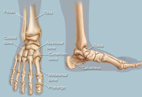
There are a variety of anatomical structures that make up the anatomy of the foot and ankle (Figure 1) including bones, joints, ligaments, muscles, tendons, and nerves. These will be reviewed in the sections of this chapter.
Regions of the Foot
The foot is traditionally divided into three regions: the hindfoot, the midfoot, and the forefoot (Figure 2). Additionally, the lower leg often refers to the area between the knee and the ankle and this area is critical to the functioning of the foot.
The Hindfoot begins at the ankle joint and stops at the transverse tarsal joint (a combination of the talonavicular and calcaneal-cuboid joints). The bones of the hindfoot are the talus and the calcaneus.
The Midfoot begins at the transverse tarsal joint and ends where the metatarsals begin --at the tarsometatarsal (TMT) joint. While the midfoot has several more joints than the hindfoot, these joints have limited mobility. The five bones of the midfoot comprise the navicular, cuboid, and the three cuneiforms (medial, middle, and lateral).
The Forefoot is composed of the metatarsals, phalanges, and sesamoids. The bones that make up the forefoot are those that are last to leave the ground during walking. There are twenty-one bones in the forefoot: five metatarsals, fourteen phalanges, and two sesamoids. The great toe has only a proximal and distal phalanx, but the four lesser toes each have proximal, middle, and distal phalanges, which are much small than those of the great toe. There are two sesamoid bones embedded in the flexor hallucis brevis tendons that sit under the first metatarsal at the level of the great toe joint (1st metatarsophalangeal joint).